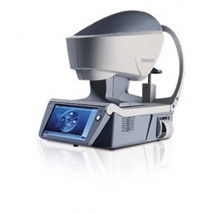
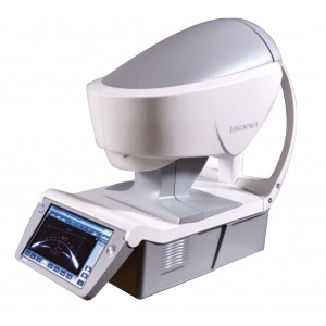
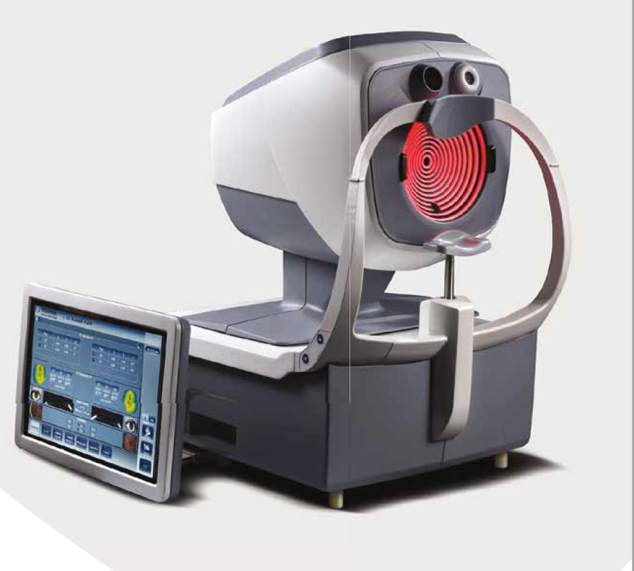
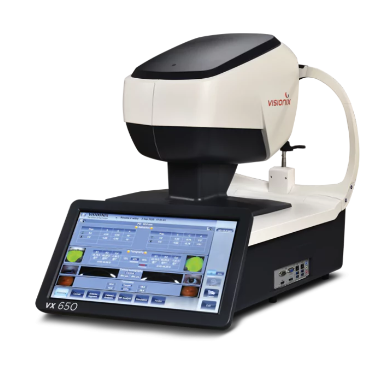
VX650
Visionix® VX650: a single multi-modal instrument for complete detection and follow-up of major anterior and
posterior ocular pathologies.
The Visionix® VX650 revolutionizes ocular assessment by introducing the first and only solution allowing eye care professionals (ECPs) to deliver a comprehensive eye exam at the push of a button.
It combines an aberrometer, a fundus camera, and all essential technologies to monitor both anterior and posterior segments in a single device. The highly automated Visionix® VX650 allows a moderately trained user to detect a wide range of visual pathologies.
-
Multi-modal device for anterior/posterior segments measurement
-
ARK
-
Aberrometer
-
Fundus camera
-
Topographer
-
Scheimpflug camera
-
Tonometer
Details
- DimensionsWIDTH 660mm
DEPTH 420mm
HEIGHT 560mm
WEIGHT 32Kg
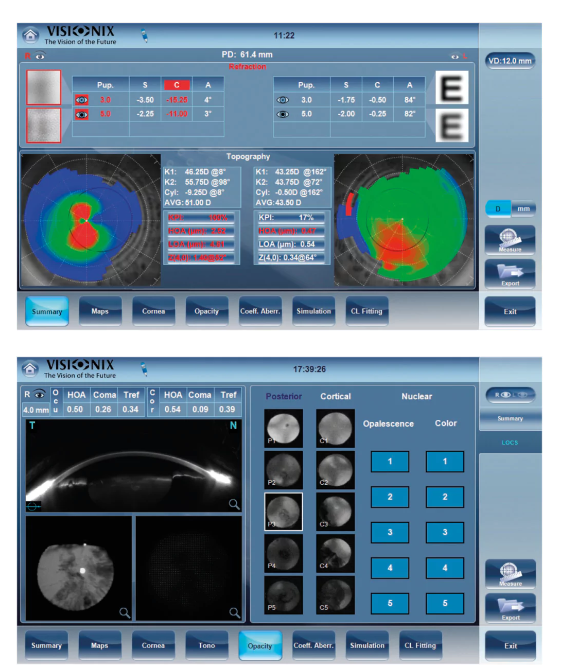
ANTERIOR SEGMENT -EYE Transparency and shape
Diagnose, Evaluate, Monitor Cataracts
• Visualization of lens opacities
• Corneal, Internal and wavefront analysis
• Internal astigmatism measurement
• Kappa angle for IOL centering
• Z.4.0 value for aspheric implant
• Lens opacity classification (LOC III scale)
ANTERIOR SEGMENT - CORNEAL TOPOGRAPHY
Diagnose, Evaluate, Monitor
Keratoconus maps
• Axial, tangential elevation and refraction maps
• Keratoconus probability index (KPI)
• Keratoconus monitoring
• Internal astigmatism measurement
• Eccentricity and meridian tables
• Corneal aberrometry
ANTERIOR SEGMENT - ADVANCED OBJECTIVE REFRACTION
Highlights differences between day and night vision needs
• Objective day and night refraction measurements
• 1400 points analysed for a 7-mm diameter pupil
• Objective refraction under mesopic and photopic conditions
• Measures lower-order and higher-order aberrations
• Access visual acuity and quality of vision on pupil as small as 1.2 mm
• Modulation Transfer Function curve analysis and comparison
Shack-Hartmann wavefront maps point out lower-order and higher-order aberrations.
ANTERIOR SEGMENT - DRY EYE DISEASES (D.E.D.)
Diagnose, Evaluate, Monitor
Displays a colour image of the eye and uses the Efron grading scale to grade the level of redness,
the overall quality of the eye and lids. This feature focuses on the meibomian glands area to screen
Meibomian Gland Dysfunction (MGD).
Using the manual zoom of the colour camera, youcan measure the height of the tear meniscus to
complete the test.
Follows the recommendations from the Tear Film& Ocular Surface Society (TFOS) and Dry Eye
Workshop (DEWS II) reports.Placido rings analysis and anterior eye camera and displays colour image of Meibomian glands.
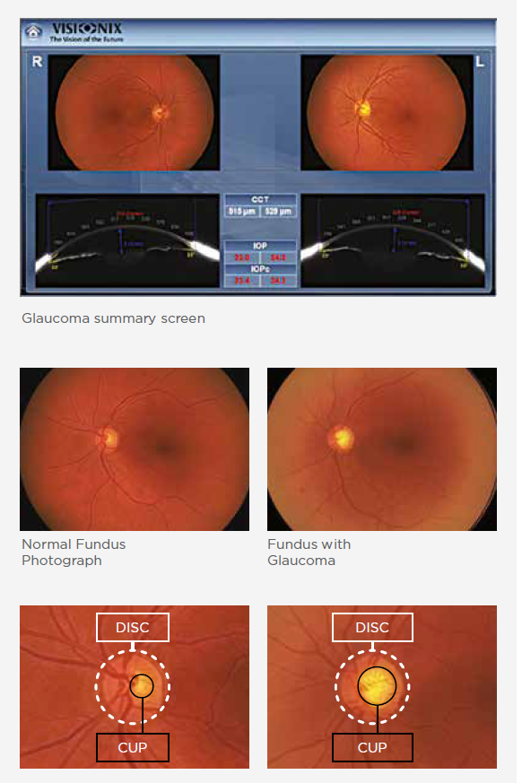
POSTERIOR SEGMENT - Glaucoma
Diagnose, Evaluate, Monitor Irido angle < 20°
IOPc
Fundus and Cup/Disc ratio
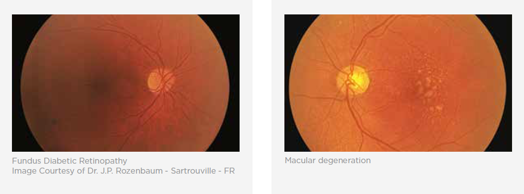
POSTERIOR SEGMENT - DIABETIC RETINOPATHY
Diabetic retinopathy can lead to other serious eye conditions:
Over time, about half of patients with diabetic retinopathy will develop Diabetic macular edemea
(DME). DME happens when blood vessels in the retina leak fluid, causing swelling in the macula
(a part of the retina). If a patient has DME, their vision will become blurry because of the extra
fluid in their macula.
An eye care professional will look at the retina for early signs of the disease, such as:
• Leaking blood vessels,
• Retinal swelling, such as macular edema,
• Pale, fatty deposits on the retina (exudates) - signs of leaking blood vessels,
• Damaged nerve tissue (neuropathy),
• Any changes in the blood vessels.
POSTERIOR SEGMENT - AMD
Diagnose, Evaluate, Monitor
Age-related Macular Degeneration is caused by the deterioration of the central
portion of the retina, the inside back layer of the eye that records the images we see and sends
them, via the optic nerve, from the eye to the brain.
The retina’s central portion, known as the macula, is responsible for focusing central vision in the
eye, and it controls our ability to read, drive a car, recognize faces or colors, and see objects in fine
detail.
Visualizing and tracking changes in retinal layers is important in managing patient visual health.